Background
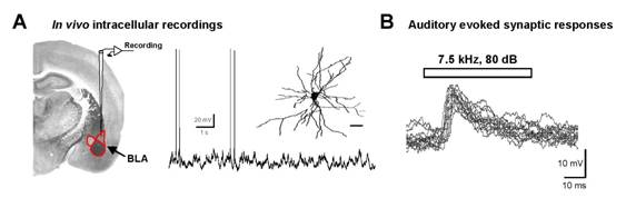 |
| Fig. 1 In vivo intracellular recordings and auditory stimulation of mouse amygdala neurons. |
A: Intracellular recordings from mouse basolateral amygdala (BLA) neurons. Left: Scheme of vertical approach of microelectrode to the BLA. Center: Spontaneous activity of a BLA neuron at rest. The cell was recorded for 2,5 h. Inset: Neurolucida reconstruction of the same neuron after revelation of Neurobiotin filling. B: Auditory stimulation via earphones inserted into the contralateral ear evokes sub-threshold short-latency postsynaptic potentials. Sweeps represent superimposed responses from 14 stimulations (repeated at 0.1 Hz).
|